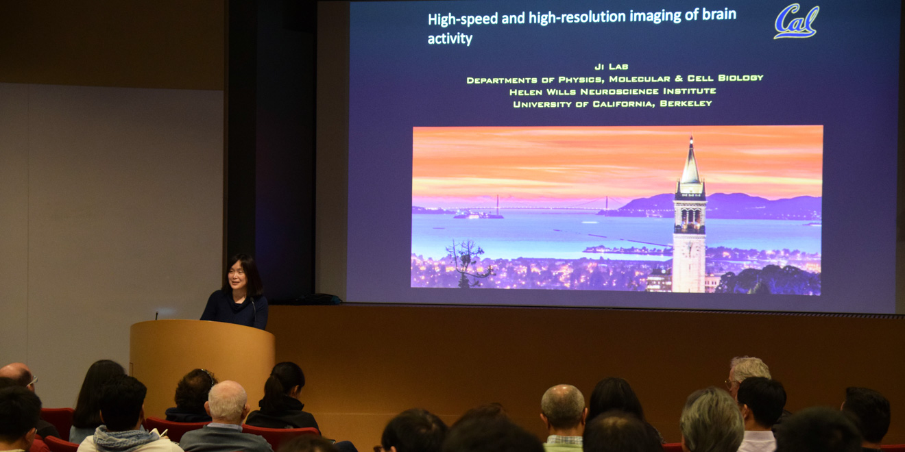
New high-speed and high-resolution imaging methods allow researchers to visualize neuronal activity in the brain on a scale that was previously unimaginable, University of California, Berkeley professor Na Ji discussed on Thursday.
To comprehend the brain, Ji explained, one needs to understand the “input-output” relationship of individual neurons — cells which are responsible for signal transmission in the nervous system, such as in the brain and spinal cord. This involves knowing what electrical information a neuron receives, and subsequently, what the neuron outputs to the rest of the brain. Such a process is the basis for interpreting neural circuits, which are populations of interconnected neurons that carry out a particular function when activated.
Recently, optical microscopy techniques have emerged as an ideal tool to help visualize the brain by giving scientists and researchers the ability to record neuron activity on a very small scale with extremely high resolution. A particular benchmark for excellent brain microscopy, Ji said, is the ability to reach sub-micron resolution in order to study nerve cell processes at depth. For size reference, a human hair is about 75 microns in diameter.
“In the past 10 years, almost, my lab has been focusing on developing methods … to achieve such high-resolution and high-speed imaging,” Ji said.
Using the mouse primary visual cortex — a region of the mouse brain that is responsible for vision — as a model system, Ji’s team drew from concepts in astronomy and optics to develop next-generation microscopy techniques to better image neural circuits with greater depth, faster speed and better resolution.
Specifically, Ji works on developing adaptive optics, a microscopy technique which deforms a mirror to reduce the effect of sample distortions on image quality. This method is typically used in astronomy to produce clear images of space.
In the biological context, this technique can be extremely helpful for visualizing individual synapses, or connections between neurons. In fact, with the aid of adaptive optics, researchers can gain novel insights into neural circuits and other brain pathways on a sub-micron level that were never before possible. For example, scientists can delve many microns deeper into brain tissue with much higher accuracy, enabling them to monitor individual neurons over multiple days to assess how the neuron is aging.
“We’re very excited,” Ji said. “We think we really have a way to do this kind of synaptic sub-cellular resolution imaging as deep as we can get.”
Contact Tejas Athni at tathni ‘at’ stanford.edu.
