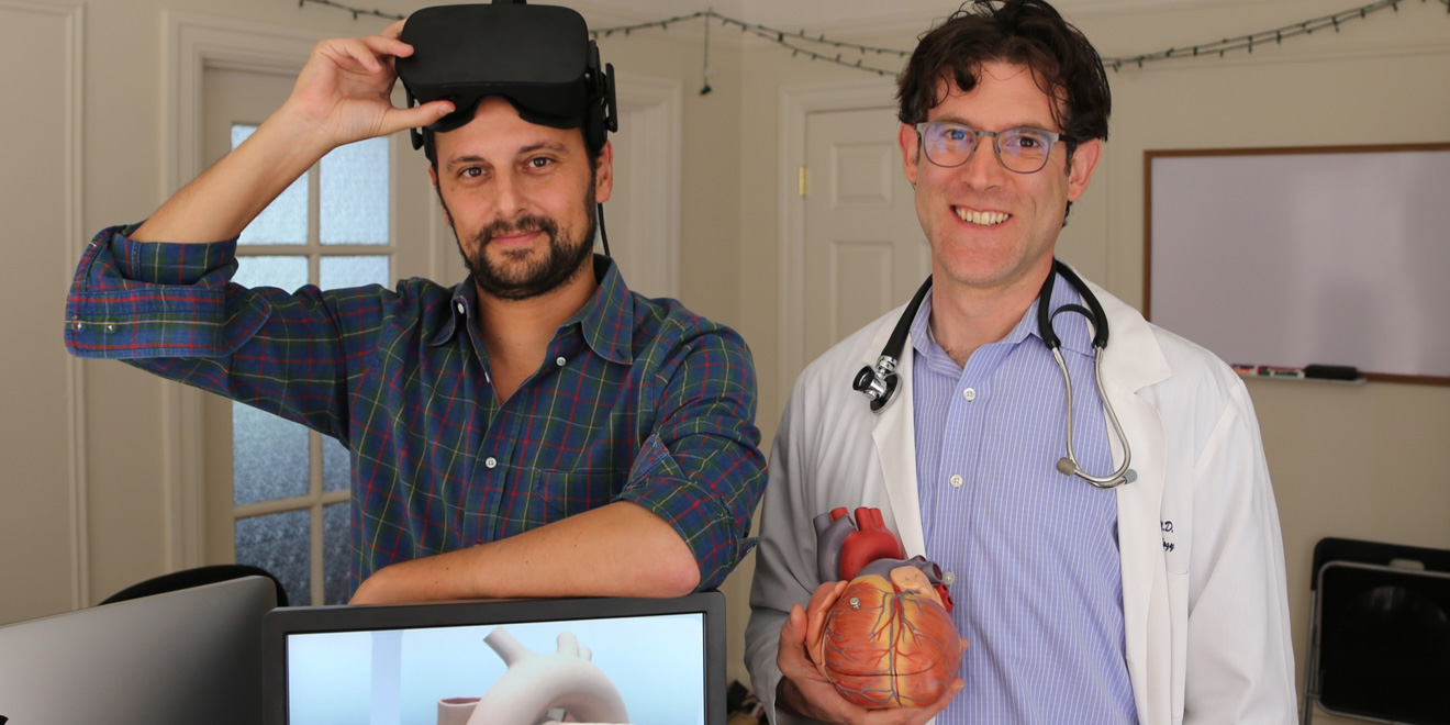
Stanford Virtual Heart, a new initiative at Lucile Packard Children’s Hospital Stanford, employs immersive virtual reality (VR) technology to tackle previously unaddressed questions in science education. Spearheaded by the hospital’s pediatric cardiologist David Axelrod, the pilot educates physician assistant students and pediatric cardiology fellows at the Stanford Medical School on congenital heart defects.
The formation of the heart is a complex process, and deviations from normal development can lead to a family of cardiac abnormalities known as congenital heart defects. Previous approaches to teaching congenital heart disease, however, were limited to plastic models, two-dimensional drawings in textbook pages and the use of cadavers, none of which can fully illustrate the dynamic, three-dimensional (3-D) nature of the heart.
The Virtual Heart’s precursor was an interactive iPad app developed in 2014 by Lighthaus, an interactive 3-D storytelling company in collaboration with Lucile Packard. The app sought to help medical trainees, families and patients better understand congenital heart disease. Its founder David Sarno — a 2013 Fellow at the John S. Knight Journalism Fellowship at Stanford — started the company during his fellowship to “use interactive graphics and virtual reality in education and storytelling.”
The project’s iPad and web-based animation allowed users to compare a healthy heart to one with a complex congenital condition called “tetralogy of fallot with pulmonary atresia and major aortopulmonary collateral arteries (MAPCAs).” Users could use the animation to visualize unifocalization, a surgical technique used to help treat MAPCA.
Earlier last year, after hearing about Lighthaus’ interactive 3-D iPad app, Axelrod reached out to Sarno to ask if he’d be interested in taking the use of technology within medical education to the next level.
“We got together and started brainstorming and thought we really ought to do all the congenital heart defects that we can in virtual reality, to try and get them all into the most immersive, interactive format that we can,” Axelrod said.
They developed the project with support with Stephen Roth, division chief of pediatric cardiology at the Betty Irene Moore Children’s Heart Center, the Lucile Packard Foundation for Children’s Health and Oculus VR.
The team consists of doctors and pediatric specialists who elucidate and define congenital heart defects, as well as software designers and animators from Lighthaus who help animate these medical conditions in the 3-D plane. Meanwhile, Oculus provides the hardware and rifts used as headsets for the project.
Equipped with a VR headset, users may choose to explore any of the 12 congenital heart defects displayed on a three by four paneled shelf. This atlas of congenital heart defects encompasses a broad spectrum of complexities ranging from a hole in the heart to defects resulting from complex biological abnormalities. When the user makes the selection, a beating heart is pulled from the shelf and projected before him. He can spin the heart around, view it from all angles, observe the vasculature, choose a certain section of the heart to observe in greater detail or even jump inside one of the heart’s chambers.
The user also may choose to “explode” the heart, causing individual components of the heart to expand away from one another such that the interior and exterior may be viewed simultaneously. This feature is Axelrod’s favorite, he said.
“It shows you what’s called the relational anatomy, what structure is next to what — and that’s something that can be really difficult to explain to people in 2-D,” Axelrod said. “That’s something that you can’t really do in other modalities.”
Axelrod and Sarno have taken the Virtual Heart prototype to a variety of recent conferences, including the Pediatric Cardiac Intensive Care Society’s international conference in Dec. 2016, international computer expo CeBIT in Mar. 2017 and Stanford Medicine X last week. Through attending these conferences, Axelrod and Sarno have received positive feedback and support from institutions, both in the US and abroad, that want to use the pilot to educate their medical trainees, they said.
“When we took [the Virtual Heart] to Germany, one of the coolest things that a number of people said, after using the demo, was ‘Oh jeez, if I’d seen this when I was in high school, maybe I would’ve wanted to go to med[ical] school,’” Axelrod said. “And I thought that was the biggest compliment ever, because you basically just told me this changed your perspective and how you would have learned biological systems. That, I think, is the gap that [VR] fills.”
The team plans to develop a full atlas of about 25 congenital heart defects that can be used in the Stanford Medical School’s residency training and advanced fellowship training programs.
But Axelrod hopes to extend the Virtual Heart’s applications further.
“We’re looking to use it certainly in the medical school, [but also] beyond — in undergraduate education in biology,” Axelrod said. “I’d also love to get it into science museums to show how the heart works.”
Looking forward, the project may also explore expanding to different medical conditions, ranging from adult heart problems like coronary issues and muscle failure to projects for a virtual brain or virtual body.
“We would really be able to focus on the immersion and interactivity [to help] people experience and learn,” Axelrod said. “The storytelling of the amazing science that goes on in all of our own bodies is what’s really the most attractive and engaging.”
Contact Katie Gu at katiegu ‘at’ stanford.edu.
